 (309) 589-1880
 ilretinst@comcast.net
(309) 589-1880
ilretinst@comcast.net
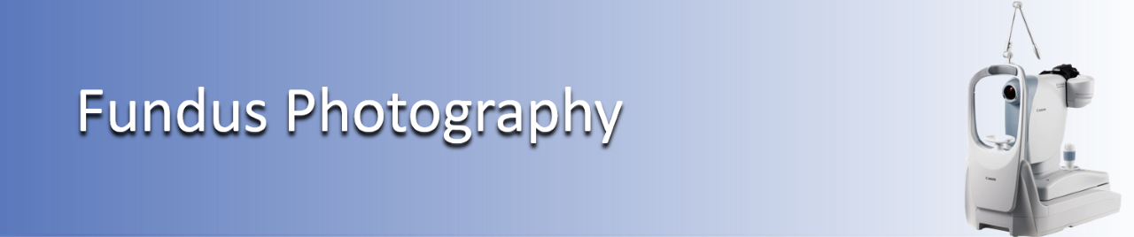
One way for our physicians to diagnose disease in the back of the eye is to take photos and do fluorescein angiography. A fundus photo is a special camera that can take pictures of the back of the eye. Sometimes the photos are done alone and often times they are done with another type of “picture” called angiography. Most people are familiar with angiography being used on the heart. We use the same idea to view the blood vessels in the back of the eye. A vegetable dye is injected into a vein in the arm or hand and as it travels through the eye, pictures are taken at different times to allow the physician to see blockages or leakage. This helps diagnosing, deciding treatments and to see if treatments are working.


 Connect with us